Respiratory Breathing System Body Organ Anatomy Diagram Vector Drawing 2181675 Vector Art at
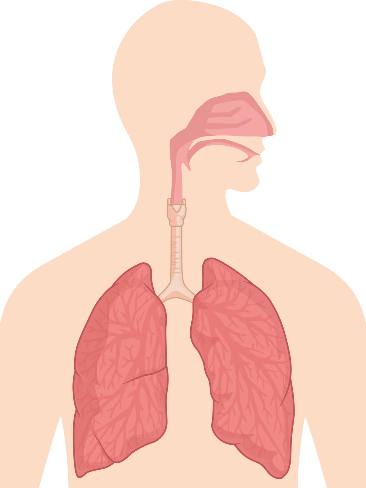
The upper respiratory tract refers to the parts of the respiratory system that lie outside the thorax, more specifically above the cricoid cartilage and vocal cords. It includes the nasal cavity, paranasal sinuses, pharynx and the superior portion of the larynx.

Human Respiratory System Vector Illustration Stock Illustration Download Image Now iStock
How to draw diagram of human Respiratory system easily - step by step Perhaps Bidesh 61.8K subscribers Subscribe Subscribed 12K 1.1M views 4 years ago Biology diagram Hi guys.. Today I will.

Respiratory System Drawing at Explore collection of Respiratory System Drawing
The respiratory system. The process of physiological respiration includes two major parts: external respiration and internal respiration. External respiration, also known as breathing, involves both bringing air into the lungs (inhalation) and releasing air to the atmosphere (exhalation). During internal respiration, oxygen and carbon dioxide.
How to draw a Human Respiratory system Diagram Drawing easy science project making step by
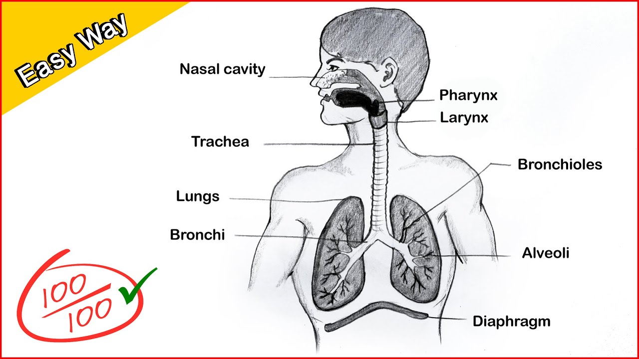
This video tutorial shows how to draw human respiratory system, in the easiest way, with step by step instructions with labeling. Its a common topic asked in CBSE & ICSE board exams and is part.
How to draw human respiratory system in easy way step by step YouTube
The respiratory system includes the organs, tissues, and muscles that help you breathe. It helps distribute oxygen throughout your body while filtering out carbon dioxide and other waste.

The Respiratory System EduPrimary
Inhalation and Exhalation. The respiratory system is the chain of organs and tissues that helps living beings breathe properly. It includes the nasal passage, mouth, lungs, and blood vessels. The muscles responsible for the contraction and relaxation of the lungs are also a part of the respiratory system.

Schematic of the human respiratory system (Adapted from [43]). Download Scientific Diagram
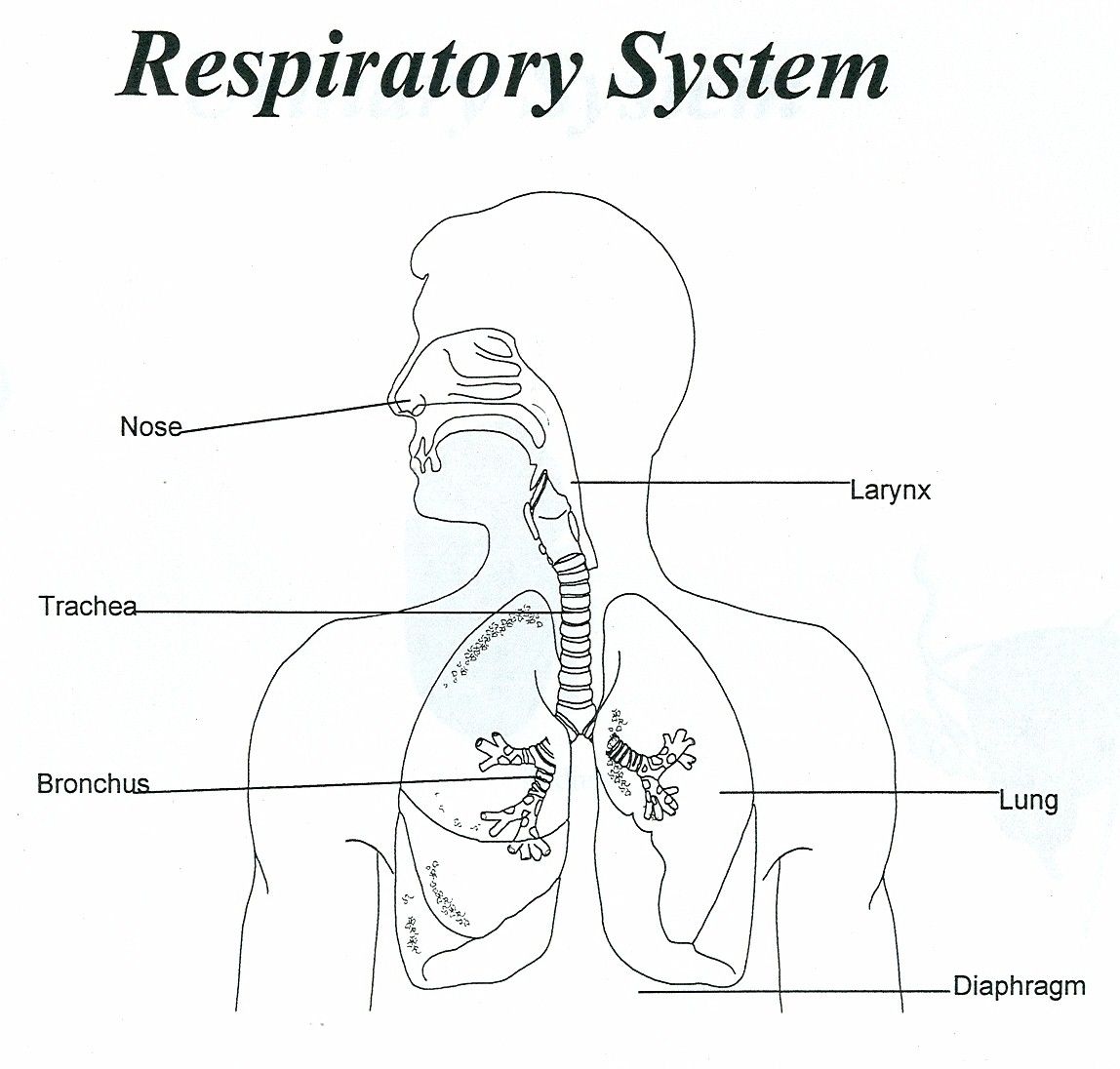
The organs of the respiratory system form a continuous system of passages called the respiratory tract, through which air flows into and out of the body. The respiratory tract has two major divisions: the upper respiratory tract and the lower respiratory tract. The organs in each division are shown in Figure 16.2.2 16.2.

Draw neat and well labelled diagram of Human respiratory system. And eplain about it Brainly.in
human respiratory system, the system in humans that takes up oxygen and expels carbon dioxide. The design of the respiratory system Passage of air through the respiratory tract explained The respiratory tract conveys air from the mouth and nose to the lungs, where oxygen and carbon dioxide are exchanged between the alveoli and the capillaries.
Respiratory System Coloring Page Coloring Home
The human respiratory system has the following main structures - Nose, mouth, pharynx, larynx, trachea, bronchi, and lungs. Explore in detail. Table of Contents Definition What Is Respiratory System Diagram Features Parts Respiratory Tract Functions Respiratory System Definition
Respiratory System Coloring Page Coloring Home
Respiratory zone. The respiratory zone, which includes the respiratory bronchioles, alveolar ducts, alveolar sacs, and alveoli, is the only site of gas exchange. Conducting zone structures. All other respiratory passages are conducting zone structures that serve as conduits to and from the respiratory zone. Stroma.
Respiratory System With Label Drawing at GetDrawings Free download
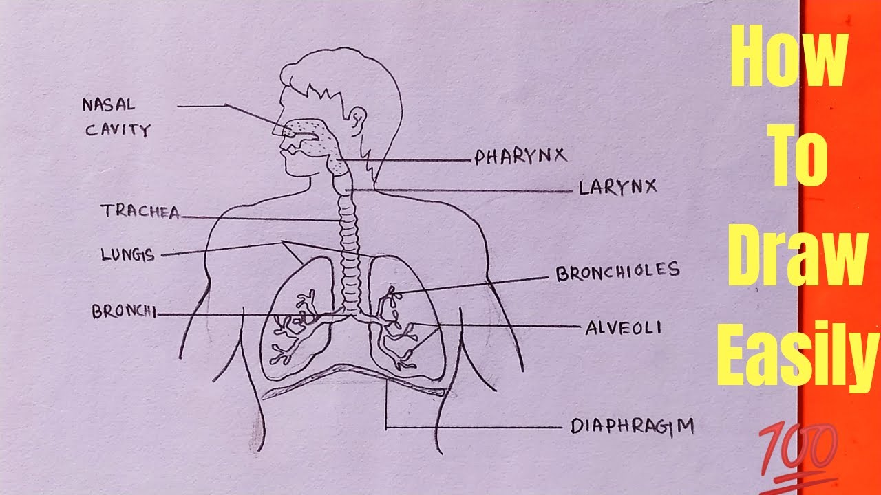
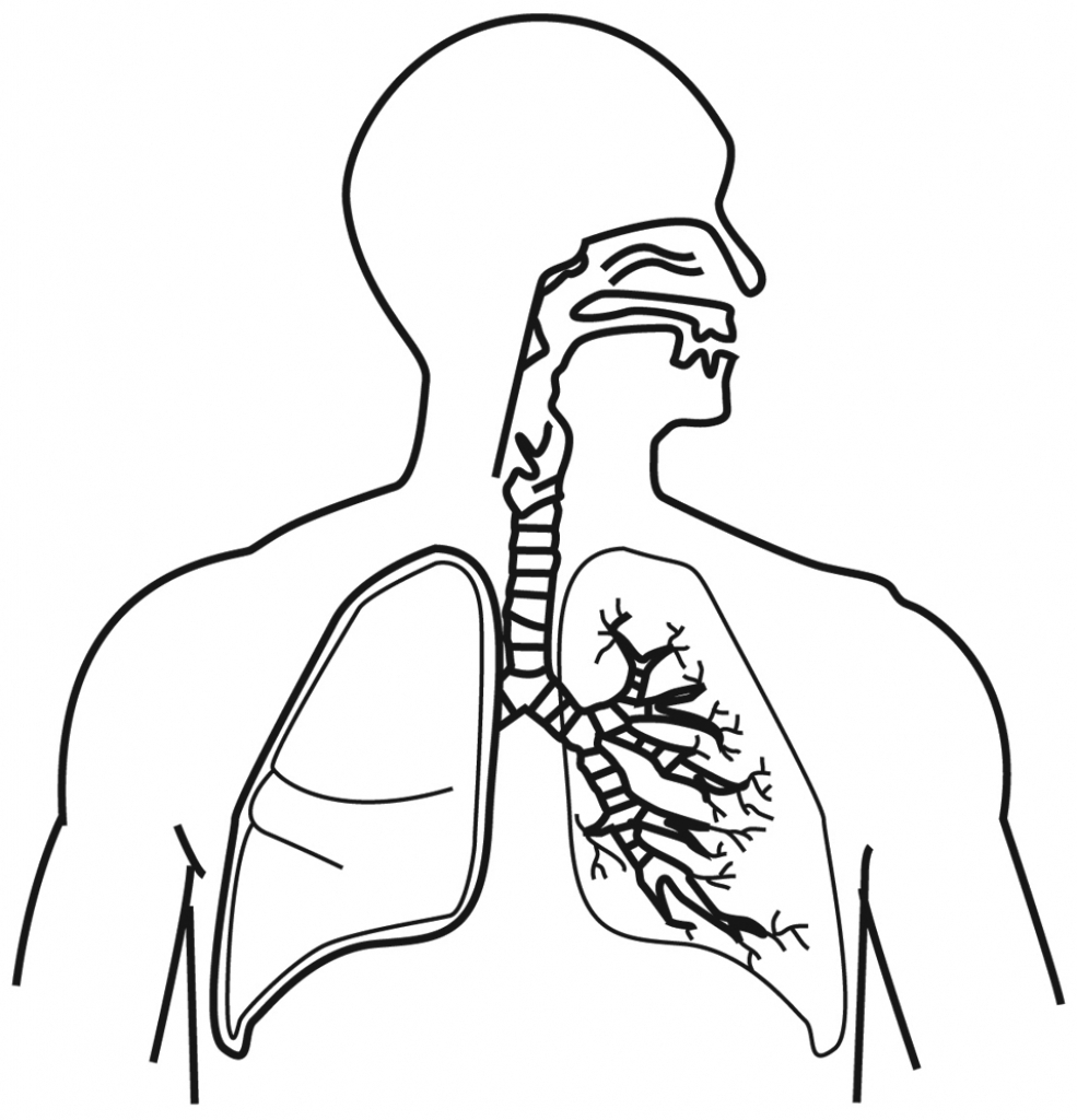
This guide will provide a description and example of how to draw the human respiratory system. First by outlining the lungs, bronchi, and trachea, then detailing additional components and accessory structures. By the end of this wikiHow, you will be proficient in drawing a medically accurate human respiratory system and be ready to ace your exams!

Respiratory System Coloring Page CC3 Classical Homeschooling for Nursing/school
Alveoli are connected to their neighbors by alveolar pores, which help maintain equal air pressure throughout the alveoli and lung ( Figure 22.11 ). Figure 22.11 Structures of the Respiratory Zone (a) The alveolus is responsible for gas exchange. (b) A micrograph shows the alveolar structures within lung tissue.

A drawing of respiratory system Brainly.in
Human Respiratory system Diagram step-by-step Drawing#abhishekdrawing #humanrespiratorysystem #biologydiagram #biology #respiratorysystem

draw a labelled diagram showing the human respiratory system Brainly.in
In the respiratory portion of the tract is connected to the bronchioles by alveolar ducts which in turn connect to alveoli. These are lined with simple squamosal epithelial. Unlike other epithelial tissue, the basement membrane of the tissue is connected to other squamosal epithelial cells; either other alveoli or capillaries. Figure 20.6. 20.6.

S04E06 How to draw Human respiratory system diagram biology YouTube
The respiratory system is the network of organs and tissues that help you breathe. It includes your airways, lungs and blood vessels. The muscles that power your lungs are also part of the respiratory system. These parts work together to move oxygen throughout the body and clean out waste gases like carbon dioxide. Advertisement.

draw a neat diagram of human respiratory system. what is the differnce between respiration amd
In the 4-week-old human embryo the beginnings of the respiratory system are first seen as an outpouching, the laryngotracheal bud, on the ventral surface of the endoderm of the digestive tract (Fig. 2.16). As the bud elongates the proximal portion forms the trachea and the distal end bifurcates to form first the two main bronchi and then the more distal parts of the bronchial tree, eventually.