
фасции шеи fascia cervicalis Анатомия, Тело, Массаж
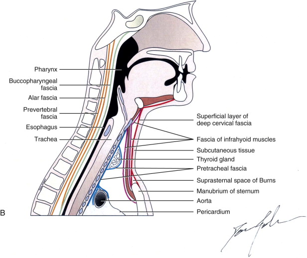
The intervening suprasternal space (of Burns) contains the lower portion of the anterior jugular veins and their connecting branch, the jugular venous arch. Four tubes are located within this superficial tube. Two of these tubes, the carotid sheaths, are paired. The carotid sheaths contain the vagus nerve, carotid artery complex, and internal.
VERTEBRAL COLUMN RIBS STERNUM by Isabella Kung Kaan
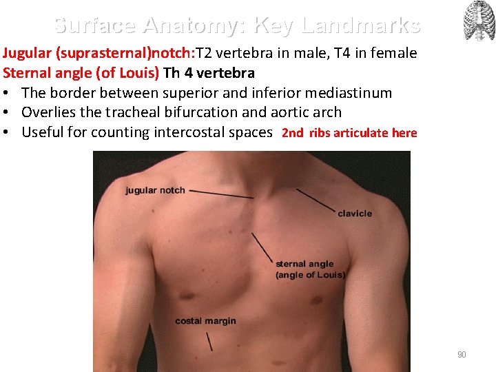
The suprasternal space (of Burns) is a space of the inferior neck. Gross anatomy. Inferior to the hyoid bone, the superficial or investing layer of the deep cervical fascia divides into anterior and posterior leafs to attach to the respective borders of the suprasternal (jugular) notch, forming a small space ~2 cm superior to the manubrium 1-3.
Ludwig’s Angina Pocket Dentistry
The suprasternal space (of Burns) is a space of the inferior neck. Gross anatomy. Inferior to the hyoid bone, the superficial or investing layer of the deep cervical fascia divides into anterior and posterior leafs to attach to the respective borders of the suprasternal (jugular) notch, forming a small space ~2 cm superior to the manubrium.
PPT WINDSOR UNIVERSITY SCHOOL OF MEDICINE PowerPoint Presentation, free download ID2716954
Suprasternal notch. Bottom of the neck; above the manubrium of the sternum, and between the two clavicles. The suprasternal notch, also known as the fossa jugularis sternalis, jugular notch, or Plender gap, is a large, visible dip in between the neck in humans, between the clavicles, and above the manubrium of the sternum .
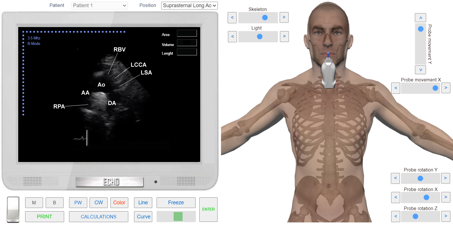
Suprasternal View. Long axis of aorta
The suprasternal space, which is also known as the 'Burns space', is a narrow space between the superficial and deep layers of the investing layers of the deep cervical fascia above the manubrium of the sternum (Fig. 1).According to Gray's anatomy, it contains a small amount of areolar tissue, the lower parts of the anterior jugular veins and the jugular venous arch, as well as the.

Echocardiographic view ; Suprasternal notch suprasternal long axis YouTube
The two spaces formed by this fascia are the posterior triangular space on the same side and the "suprasternal space of Burns" in the midline. The suprasternal space arises when two layers of fascia attach to the front and back of the sternum. middle layer-visceral layer;

Suprasternal notch Anatomical Terms Pronunciation by Kenhub YouTube
This creates the variably sized suprasternal space of Burns (or Gruber), which contains fat and a communicating vein between the left and right anterior jugular veins. If this space is entered during a tracheostomy, inadvertent transection of the communicating vein may result in considerable blood loss. Caudally the SLDCF is attached into the.

Tuberculous suprasternal notch abscess in a child Vijay and Vaishya 2016 BMJ Case Reports
Anterior triangle of the neck. Covers. Infrahyoid muscles. Suprasternal space (of Burns) In the Suprasternal notch, the investing fascia splits into two layers: One attaches to the anterior border of the manubrium. The other to the posterior border. This leaves a small suprasternal space containing: A little fat.
Investing layer of deep cervical fascia DETAILED (1/4) YouTube
The suprasternal space (Space of burns) is a narrow space between the superficial and deep layers of the investing layers of the deep cervical fascia.. Boundaries: Anterior: superficial layer of deep cervical fascia attached to the anterior border of the manubrium; Posterior: deep layer of deep cervical fascia attached to the posterior border of manubrium and to the interclavicular ligament.

A) The suprasternal twodimensional echocardiographic view of the... Download Scientific Diagram
The suprasternal space is a narrow space between the superficial and deep layers of the investing. layers of the deep cervical fascia above the manubrium of the sternum. The suprasternal space has. been paid little attention as a space with the potential for lymph node metastasis from both thyroid. cancer and head and neck cancer.
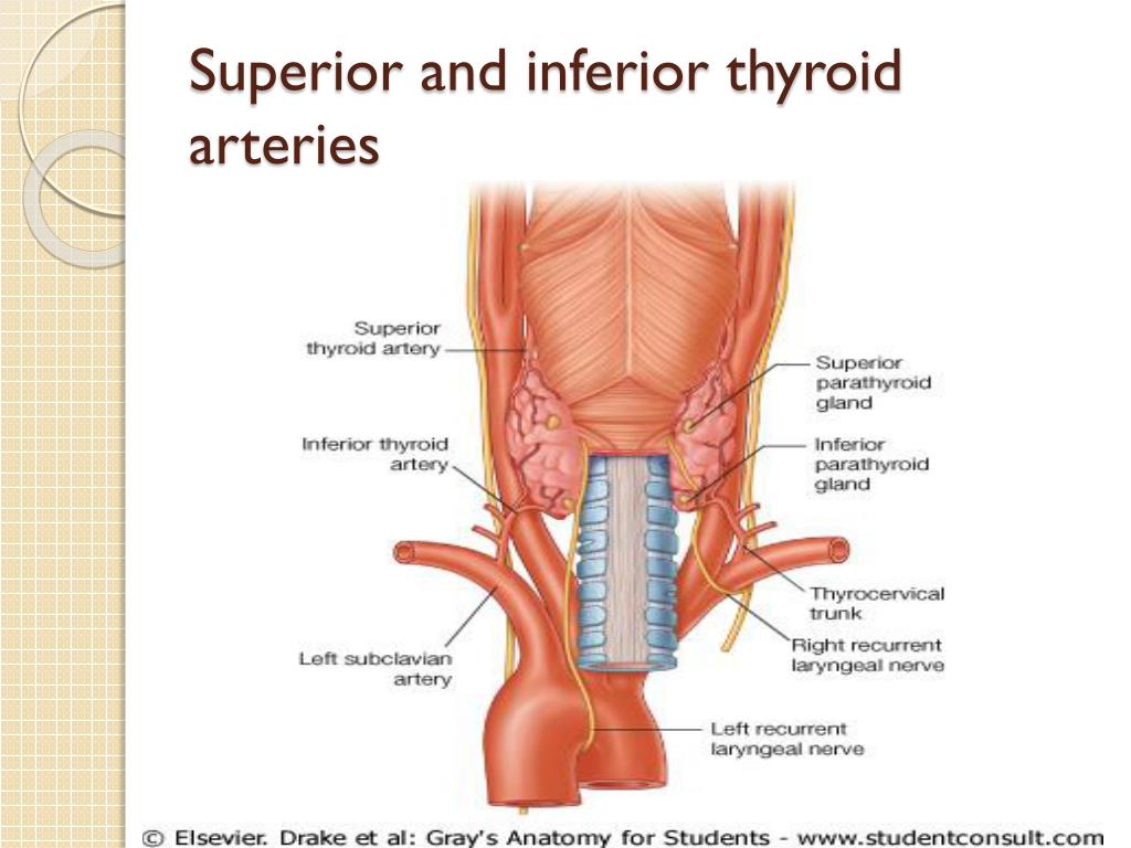
Esophageal branches of inferior thyroid artery (Rami oesophageales arteriae thyroideae
The suprasternal space, also called the space of Burns, consists of superficial and deep layers of the investing layers of the deep cervical fascia above the manubrium of the sternum [10]. It has little areolar tissue and few lymph nodes.

PPT Understanding the Fascial planes of head and Neck PowerPoint Presentation ID6031589
A. Rule of 7s for Duration of Swelling: If 7 days: Inflammatory. If 7 months: Neoplastic. If 7 years: Congenital. B. 80:20 rule for Malignant vs Benign Neck Swellings: In Pediatric age group: 20% are Malignant and 80% are Benign. In Adult age group: 20% are Benign and 80% are Malignant. C. 20:40 rule for Age group: <20 years:

Tuberculous suprasternal notch abscess in a child BMJ Case Reports
The suprasternal space of Burns (a suprasternal midline space created by a leaflet split in the superficial layer of deep fascia) is opened while leaving the periosteum and anterior transthoracic fascia for protection of the brachiocephalic veins. Next, the omohyoid muscle is marked and divided prior to identifying the internal jugular vein and.

Bildergebnis für fascia Deep fascia, Subcutaneous tissue, Fascia
LNCM space includes suprasternal space and intra-infrahyoid strap muscle space.. (LNSS) in lateral neck dissection , which anatomically classified as part of the space of Burns. They concluded that the positive rate of LNSS was 22.6% in clinically node-positive (cN+) PTC, which was correlated with a primary site in the inferior pole,.

2a Horizontal CT view of the neck at the level of suprasternal notch;... Download Scientific
suprasternal space of Burns in the midline. The middle layer of the deep cervical fascia is also known as the visceral fascia It has two subdivisions, the muscular division, which surrounds the infrahyoid strap muscles, and the visceral division, which envelops the pharynx, larynx,

Αποτέλεσμα εικόνας για suprasternal notch Medical anatomy, Anatomy images, Anatomy
The suprasternal space, also called the space of Burns, consists of superficial and deep layers of the investing layers of the deep cervical fascia above the manubrium of the sternum [10].. The rate of lymph node metastasis of thyroid or head and neck cancer in suprasternal space has been investigated by few studies [3,4]. In their.